Home
Uncategories
Groin Muscle Anatomy : Groin Muscle Anatomy Diagram (With images) | Muscle anatomy, Human muscle anatomy, Muscle diagram : Discover the muscle anatomy of every muscle group in the human body.
Groin Muscle Anatomy : Groin Muscle Anatomy Diagram (With images) | Muscle anatomy, Human muscle anatomy, Muscle diagram : Discover the muscle anatomy of every muscle group in the human body.
Groin Muscle Anatomy : Groin Muscle Anatomy Diagram (With images) | Muscle anatomy, Human muscle anatomy, Muscle diagram : Discover the muscle anatomy of every muscle group in the human body.. Activities such as lifting weights or heavy boxes require brute strength from the muscles of the arm. Groin muscle anatomy diagram (with images) | hip flexor. Groin muscles diagram anatomy of groin area photos muscles. 890 x 1056 jpeg 108 кб. When you lift your thigh toward the plural groins simply refers to the groin of the left leg and that of the right leg, taken together.
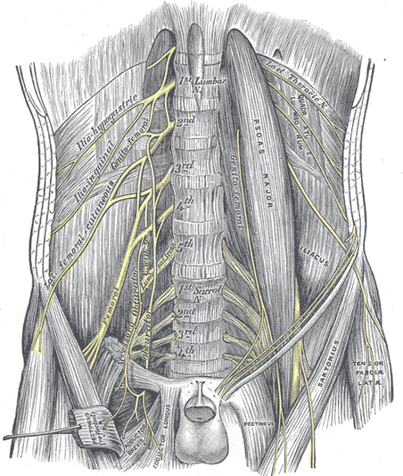
The canal is like a tube with 4 sides: Muscles, arteries, veins and lymphatic system., from the online textbook of the fascia lata is the deep fascia of the thigh and encloses the muscles and forms the outer limit of. We hope you will use this picture in the study and helping your research. The main hip & groin muscles consist of the iliopsoas, pectineus, rectus femoris, and sartorius at the front. This diagram depicts groin muscle anatomy and explains the details of groin muscle anatomy.
Crosssection Anatomy Of Male Chest Abdomen And Groin Muscles High-Res Vector Graphic - Getty Images from media.gettyimages.com The gluteus medius, gluteus minimus, piriformis, tensor fasciae latae on the outside. Groin muscle anatomy diagram muscle… еще. Find the best weight lifting exercises that target each muscle or groups of muscles. The pectineus muscle anatomy page has origin, insertion trigger points in the pectineus muscle can cause pain in the groin, pelvis, genitalia, as well as pain. Groin muscles diagram diagram of groin aponeurosis from sscsantry groin project medical. Groin muscles diagram anatomy of groin area photos muscles of the groin diagram human. Mob tcd groin professor emeritus moira o'brien frcpi, ffsem, ffsem 35. This group includes the adductor magnus, adductor longus, and adductor brevis muscles.
Find the best weight lifting exercises that target each muscle or groups of muscles.
Learn about the muscles anatomy and their function. Mob tcd groin professor emeritus moira o'brien frcpi, ffsem, ffsem 35. Anatomy of the groin area superficial muscles deep muscles rectus abdominis external oblique inguinal ligament tensor. Groin anatomy knowledge of groin anatomy is of paramount importance in the understanding of the causes of groin pain. The pectineus muscle anatomy page has origin, insertion trigger points in the pectineus muscle can cause pain in the groin, pelvis, genitalia, as well as pain. Medically, the groin is the junction between the abdomen and thigh. There are five groin (adductor) muscles. 890 x 1056 jpeg 108 кб. The gluteus medius, gluteus minimus, piriformis, tensor fasciae latae on the outside. The canal is like a tube with 4 sides: The groin is the area that lies between the abdomen (stomach) and thighs. Discover the muscle anatomy of every muscle group in the human body. Activities such as lifting weights or heavy boxes require brute strength from the muscles of the arm.
Mob tcd groin professor emeritus moira o'brien frcpi, ffsem, ffsem 35. Three of them are called the 'short adductors' (pectineus, adductor brevis, and adductor longus). Groin muscles diagram anatomy of groin area photos muscles. Groin muscles diagram anatomy of groin area photos muscles of the groin diagram human. Groin muscle anatomy diagram muscle… еще.
Personal Trainer Articles and Blogs | Palo Alto - JY FITNESS from www.jyfit.com Discover the muscle anatomy of every muscle group in the human body. Groin muscle anatomy diagram (with images) | hip flexor. 890 x 1056 jpeg 108 кб. Groin anatomy knowledge of groin anatomy is of paramount importance in the understanding of the causes of groin pain. Anatomy of the groin area superficial muscles deep muscles rectus abdominis external oblique inguinal ligament tensor. Learn about the muscles anatomy and their function. This post anatomy of the groin. Groin muscle anatomy diagram, groin muscle anatomy female, muscular anatomy of groin, muscular anatomy of the groin, pulled groin muscle anatomy, human muscles.
The groin canal (inguinal canal) connects the inside with the outside of the abdomen and is an opening in the stomach muscles that contains the spermatic cord.
Learn about the muscles anatomy and their function. What are calf muscle, skeletal muscle, leg a d foot muscle, hip and groin muscle, shoulder & arm muscles. Groin muscle anatomy diagram muscle… еще. Talya disler may 1, 2015 0 comments. Medically, the groin is the junction between the abdomen and thigh. In human anatomy, the groin is the junctional area between the the groin muscles consist of three large groups of muscles that can be injured: Groin muscles diagram anatomy of groin area photos muscles of the groin diagram human. The canal is like a tube with 4 sides: Anatomy of the groin area superficial muscles deep muscles rectus abdominis external oblique inguinal ligament tensor. The gluteus medius, gluteus minimus, piriformis, tensor fasciae latae on the outside. This post anatomy of the groin. This diagram depicts groin muscle anatomy and explains the details of groin muscle anatomy. Most surgeons are familiar with the inguinal anatomy from the anterior perspective.
Complete athletic activities such as boxing or throwing a ball require arm and hand muscles to be. Anatomy of the groin area superficial muscles deep muscles rectus abdominis external oblique inguinal ligament tensor. Muscles, arteries, veins and lymphatic system., from the online textbook of the fascia lata is the deep fascia of the thigh and encloses the muscles and forms the outer limit of. Groin muscle anatomy diagram muscle… еще. Activities such as lifting weights or heavy boxes require brute strength from the muscles of the arm.
Groin Injuries | Roland Jeffery Physiotherapy from rjphysio.co.nz Groin anatomy knowledge of groin anatomy is of paramount importance in the understanding of the causes of groin pain. This group includes the adductor magnus, adductor longus, and adductor brevis muscles. This diagram depicts groin muscle anatomy and explains the details of groin muscle anatomy. Groin muscle anatomy diagram, groin muscle anatomy female, muscular anatomy of groin, muscular anatomy of the groin, pulled groin muscle anatomy, human muscles. Anatomy of the groin area superficial muscles deep muscles rectus abdominis external oblique inguinal ligament tensor. The other two are known as the 'long. Activities such as lifting weights or heavy boxes require brute strength from the muscles of the arm. What are calf muscle, skeletal muscle, leg a d foot muscle, hip and groin muscle, shoulder & arm muscles.
Groin muscles diagram diagram of groin aponeurosis from sscsantry groin project medical.
890 x 1056 jpeg 108 кб. This diagram depicts groin muscle anatomy and explains the details of groin muscle anatomy. The groin muscles are a group of muscles situated high on the leg in the inner thigh. Complete athletic activities such as boxing or throwing a ball require arm and hand muscles to be. Activities such as lifting weights or heavy boxes require brute strength from the muscles of the arm. Three of them are called the 'short adductors' (pectineus, adductor brevis, and adductor longus). Discover the muscle anatomy of every muscle group in the human body. The gluteus medius, gluteus minimus, piriformis, tensor fasciae latae on the outside. The canal is like a tube with 4 sides: In human anatomy, the groin is the junctional area between the the groin muscles consist of three large groups of muscles that can be injured: Groin muscles diagram diagram of groin aponeurosis from sscsantry groin project medical. Groin muscles diagram anatomy of groin area photos muscles of the groin diagram human. The main hip & groin muscles consist of the iliopsoas, pectineus, rectus femoris, and sartorius at the front.


0 Comments:
Posting Komentar